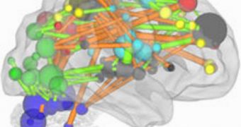
A team of investigators managed to use an established form of brain imaging to produce maps of how neural networks change and evolve in the human brain over a period of time.
Mapping out these complex interactions was no easy task, but determining this data was of the utmost importance. The study was mostly conducted on children.
Because experts now know how the brain changes, they can start looking into the cortices of children suffering from very specific conditions, such as autism.
In such studies, they may be able to determine whether “hardware” changes are behind the development of the condition, or if it is caused by glitches elsewhere in the brain.
In the near future, even more applications are conceivable. Researchers say that they could use the new method of analyzing brain images to determine which of the children that have abnormal brain structures will actually develop conditions such as autism.
The investigation was conducted by a team of experts at the Washington University School of Medicine (WUSM), Technology Review reports.
The group relied on a brain-imaging technique known as functional Magnetic Resonance Imaging (fMRI) for its study. This method provides a relatively simple way of keeping track of what goes on in the brain at any given time.
Patients or test subject simply lie on a lab table, doing nothing in particular, while the machine analyzes the way various areas of their brain fire up in sync as the people think of various things.
Though fMRI is a valuable scientific tool, it cannot be used to make diagnostics. Rather, it is used to collect data from multiple research subjects, and derive conclusions from that.
“We realized that if the promise of fMRI is going to apply to understanding individual patients, we need to come up with a new approach, one that can take advantage of an individual's data,” explains the leader of the new research, Bradley Schlaggar.
The expert is a WUSM pediatric neurologist. His team managed to develop a twist on functional connectivity, which in turn allowed the researchers to derive new sets of data from their work.
Computers were used to detect correlations and changes in the way neural networks were set up in individuals ranging in age from 5 to 30.
The complex algorithms used allows the team to find changes that would have been impossible to decipher for the naked eye.
The WUSM group says that it will continue to improve the new observations method.

 14 DAY TRIAL //