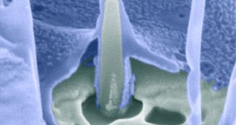
A group of physicists at the Stanford University has developed a new method for studying the insides of a cell. The approach, which will benefit neurologists, biologists and a whole bunch of other scientists, is dependent on the use of glowing nanopillars.
The novel cellular research platform features pillar-like structures at the nanoscale, which are capable of glowing in a way that enables researchers to peer inside living cells. Generally, studying living biological system is extremely complex.
Primarily, this is because existing imaging technologies in this domain are highly invasive, and tend to destroy the samples they are analyzing. Experts have been trying to develop less destructive methods of study in this area for years.
In a recent article published in the esteemed journal Proceedings of the National Academy of Sciences (PNAS), the Stanford team says that their new imaging system is tremendously precise at its job.
“The nanopillar structures themselves offer many advantages that make this development particularly promising for the study of human cells,” explains Stanford assistant professor of chemistry Bianxiao Cui, the senior author of the research.
The expert conducted the work with engineer Yi Cui and scholars Chong Xie and Lindsey Hanson. The platform relies on surveying backlight, rather shining light directly on the target sample.
In order to succeed in their effort, the researchers had to create light sources that shone at wavelengths smaller than that of visible light. A basic limitation in physics prevents experts from seeing objects smaller than 400 nanometers.
But the quartz nanopillars used in this study produce evanescent light waves that travel for only 150 nanometers in all direction. This means that the light source is smaller than the wavelength of light.
This allows experts to see objects that are two times smaller than the smallest objects visible with any other imaging method. As such, scientists will be able to image structures less than 200 nanometers in size, which was previously impossible without destroying the samples.
The reason why this ability is so important is because even the most common proteins at work in human cells are smaller than 400 nanometers. But many of them can be observed in the 200 to 400 nanometer range.
But the Stanford team also took its study one step further, and developed a method of targeting their diminutive light sources on specific structures inside cells.
“We know that proteins and their antibodies attract each other. We coat the pillars with antibodies and the proteins we want to look at are drawn right to the light source – like prima donnas to the limelight,” Bianxiao Cui explains.
“The nanopillars look a bit like tiny light sabers, but they provide just the right amount of light to allow scientists to do some pretty amazing stuff – like looking at individual molecules,” concludes Stanfrod associate professor of materials science and engineering Yi Cui.

 14 DAY TRIAL //