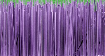
A new microscopic imaging technique makes use of the smallest nanoscale applications created by man to take snapshots of incredibly tiny biological structures, like viruses and proteins, smaller than an individual cell.
The new method would be able to perform this task with unprecedented resolution with the help of a nanowire suspended in a force field and an optical sensor behind it. The force field itself is invisible and it's made of laser radiation.
A nanowire is a wire of dimensions the size of a nanometer (a billionth of a meter). Alternatively, nanowires can be defined as structures that have a lateral size constrained to tens of nanometers or less and an unconstrained longitudinal size.
The prototype of the new imaging sensor has been developed by Peidong Yang and his colleagues at the University of California, Berkeley and works by scanning the superfine tip of the nanowire placed over a biological sample, with a laser beam that it emits from the other end of the nanowire.
The light that gets through the object is caught by an optical sensor located at the back end of the wire, which builds up a picture of the sample's structure. The geometry of nanowires, with a lot more surface area compared to volume, as the new research shows, makes them inherently good at sensing light.
Early demonstrations of the prototype proved it to have high imaging capabilities, being able to achieve a resolution of 100 nanometres, enough to capture nanoscale features, like those of a silicon chip. The creators of this new technique are confident that the resolution could be increased even further, by refining the device, so that it will eventually reach a resolution of only tens of nanometers.
Upon completion, the new device could lead to increasing usage of nanowires in photodetection and photovoltaic applications.

 14 DAY TRIAL //