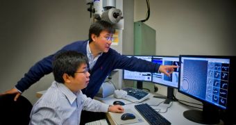
A group of experts at the U.S. Department of Energy's (DOE) Lawrence Berkeley National Laboratory (Berkeley Lab) were recently able to produce the first 3D image of an individual protein ever obtained.
This was made possible when scientists at the lab's Molecular Foundry pushed their Zeiss Libra 120 Cryo-Tem microscope far beyond the capabilities it was designed to display. The instrument itself is extremely expensive, billed at around $1.5 million.
Cryo-electron microscopes can be used to obtain images of unprecedented accuracy levels, but even they have some limitations. What researcher Gang Ren and his group did was tweak the instrument to such an extent that it became capable of revealing individual molecules in extreme detail.
Remarkably, the Molecular Foundry group is now able to observe single protein with such clarity that they can make out its structure. This is indescribably important for the field of medicine, where countless therapies and drugs act on proteins that researchers don't really know how they look like.
At this point, most of our understanding of protein structures comes from approximations. What this new observations technique will do is allow us to figure out the exact structure of such molecules directly, without interference or proxies.
Currently, experts used techniques such as X-ray diffraction, nuclear magnetic resonance, and conventional cryo-electron microscope (cryoEM) imaging to figure out how proteins look like, but these approaches work by creating an average set of data after analyzing millions of proteins.
Ren termed the new CEM technique “individual-particle electron tomography” (IPET). The way it works is described in a paper published in the January 24 issue of the peer-reviewed scientific journal PLoS ONE, which is edited by the Public Library of Science.
“The 3-D images reported in the paper include those of a single IgG antibody and apolipoprotein A-1 (ApoA-1),” the researchers say. Ren adds that the structure of “good cholesterol” molecules, called HDL, will soon be unraveled.
Numerous research teams have been trying to figure out how this molecules look like for years, but their efforts have failed despite their best efforts. Targeting HDL could help alleviate several major diseases of the cardiovascular system, therefore reducing risk factors for depression, diabetes, obesity and other major, widespread diseases.

 14 DAY TRIAL //