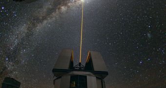
Technologies originally developed for space have been used for a host of civilian applications for a long time, and it would appear that now it's time for adaptive optics to get a new twist. The study method has recently been fine-tuned for looking inside cells, rather than at the most distant stars.
Whenever astronomers want to use ground-based telescopes to view something that is far away from Earth, they need to compensate somehow for the turbulences caused by Earth's flickering atmosphere.
The method they developed to do this is called adaptive optics. Telescopes featuring it have mirrors that are made up of segments which can move independently. When observing the night sky, these segments move slightly, in such a way that the interferences caused by the atmosphere disappear.
But, in order for the technique to work, experts need to use a powerful laser beam to create an artificial point of reference, called a guide star. Now, experts create a similar guide star for biomedical imaging.
These types of observations could from now on become considerably more accurate, if the new method is employed ad a wide scale. Behind the work was Washington University in St. Louis (WUSL) investigator Lihong Wang, PhD.
The expert, who holds an appointment as the Gene K. Beare Distinguished Professor of Biomedical Engineering at WUSL, created the artificial guide star out of an ultrasound beam.
What this beam does is it literally tags whatever light passes through it. After that light passes through the tissue sample, it is combined with a reference beam to create a hologram. A “reading beam” is then passed through the hologram.
This creates light waves that travel back through the tissue sample, and then re-focus at their source, where the ultrasound beam is also focused. What the technique does is it allows experts carrying out the measurements to control where light is focused inside a sample.
The imaging method has been named time-reversed ultrasonically encoded (TRUE) optical focusing, and its creators believe it will play an important part in future medical research and diagnostics.
“Focusing light into a scattering medium such as tissue has been a dream for years and years, since the beginning of biomedical optics. We couldn’t focus beyond say a millimeter, the width of a hair, and now you can focus wherever you wish without any invasive measure,” Wang explains.
Details of how TRUE optical focusing works have been published in the January 16 online issue of the top scientific journal Nature Photonics, and have also been highlighted in the journal Physics Today.

 14 DAY TRIAL //