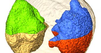
Scientists have been trying to develop a way of measuring the atomic structure of nanomaterials directly for quite some time now. During a new investigation, researchers at the University of California in Los Angeles (UCLA) may have come up with a method to do just that.
Their approach enables them to visualize individual atoms within nanoparticles, as well as map their positions in three dimensions. Details of how the imaging method functions were published in the March 22 issue of the top scientific journal Nature.
The UCLA team used a scanning transmission electron microscope to achieve its objectives. The instrument is capable of focusing a narrow beam of high-energy electrons onto very small targets, in this case a gold nanoparticle just 10 nanometers across.
Despite its small size, the nanoscale particle contains tens of thousands of individual gold atoms. The UCLA group says that each of these atoms is about one million times smaller than the average width of a human hair.
“This is the first experiment where we can directly see local structures in three dimensions at atomic-scale resolution – that's never been done before,” California NanoSystems Institute (CNSI) researcher Jianwei Miao explains. He is also a professor of physics and astronomy at UCLA.
The scientist says that the operating principle behind their technique is fairly simple. The electron beam passes through the gold sample, casting shadows on a detector located behind the microscope. This background is able to decipher data contained in the shadows individual gold atoms cast.
In order to reconstruct the 3D configuration of the nanoparticle, the scientists had to take measurements of the sample from 69 different angles, and then combine them into a single view. This method is known as electron tomography.
The technique does have a flaw, however, which is that it loses some of the information each atom carries. “It is like averaging together everyone on Earth to get an idea of what a human being looks like – you completely miss the unique characteristics of each individual,” Miao says,
“Our current technology is mainly based on crystal structures because we have ways to analyze them. But for non-crystalline structures, no direct experiments have seen atomic structures in three dimensions before,” he concludes.

 14 DAY TRIAL //