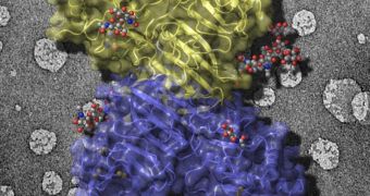
A group of investigators at the University of Michigan and Purdue University, in the United States, managed in a recent study to identify key aspects of a process through which viruses responsible for Dengue fever and West Nile fever replicated within host cells.
These results are very important because they open up new avenues of research when it comes to fighting these mosquito-borne diseases. While the two conditions have been largely eradicated in more developed parts of the world, they still represent important public health threats in many regions.
Researchers say that there is currently no vaccine for either of them. This is partially what prompted the research team to study viral replication in these two agents. The group was also able to figure out how the viruses manipulate the human immune system as they spread through the body.
Details of the investigation, which was led by U-M Life Sciences Institute expert Janet Smith, appear in a paper published in the February 6 online issue of the top journal Science. The main achievement was recreating the structure of a protein that helps the two viruses replicate and infect hosts faster.
“Seeing the design of this key protein provides a target for a potential vaccine or even a therapeutic drug,” Smith explains. The protein in question is called NS1, and is produced exclusively in infected cells. When released into the bloodstream, this molecule masks the viral agents, safeguarding them from the immune system.
This is very dangerous for patients, since both agents are part of the flavivirus family, which also includes yellow fever and several other encephalitis viruses. Though originating in the Equatorial Belt, Dengue fever even made its way to the United States, and infects around 400 million people annually.
The West Nile virus infected almost 3,000 people last year in the United States alone. These incidences could be decreased following the new study, which used X-ray crystallography to map the position of all atoms in the protein.
“Isolating the protein in order to study it has been a challenge for researchers. Once we discovered how to do that, it crystallized beautifully,” Smith concludes.

 14 DAY TRIAL //