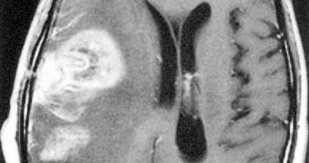
The brain may be an elastic organ, but once the skull is opened, it's not longer floating in the protective bath of cerebrospinal fluid, and gravity forces and atmospheric pressures change its shape, but also the location of the tumor(s) to be removed.
The preoperative MRI (magnetic resonance imaging) is no longer precise, and the brain's shape changes further once the surgeon starts the operation. The space issue is linked to the time one: removing as much as possible of the tumor without touching healthy neural tissue, as the slightest errors can lead to severe consequences.
The problem is that MRI requires time, and each intraoperative MRI can take as long as 90 minutes. "They tell me that they don't even talk while the MRI is happening," said Nikos Chrisochoides, a professor of computer science at the College of William and Mary in Virginia, lead researcher of a group working with a Harvard team for employing mathematics and computer power to shorten the time issue. The team would provide the surgeons with a dynamic computer model of the patient's brain.
"In clinical trials, my team can render a new model in six or seven minutes, but hopes to be able to do so in under two minutes. We want to help the neurosurgeon make an informed decision of what to cut, where the critical paths are, what areas to avoid. I'm neither a neurosurgeon nor a doctor, so the contribution of my research is to make this distillation of objects really, really, really fast.", said Chrisochoides.
The projection computer monitor has a screen similar to those found in small multiplex theaters. 3-D glasses give an amazing 3-D effect, presenting how displacement impacts the brain and the position of the tumor.
A variety of images are acquired before the surgery, and low-resolution intraoperative data permits the detection of the change in brain matter, determining modifications in the preoperative images according to them.
"If you push (the brain), it takes energy and then after a while it settles down. We can calculate the place where it settles by solving the partial differential equation. Mathematicians can tell us that there is a solution, but they cannot tell us what the solution is. There's no such thing for this equation. There's no analytic solution. So we have to approximate.", said Chrisochoides.
The approximation of the brain's geometry is made by tessellation: division in triangles in 3-D (a mesh representation of the brain). "This is a great example of how computer sciences research is impacting other fields and enables such important capabilities, and it's really great to see this impact in medicine.", said Frederica Darema, a NSF officer.

 14 DAY TRIAL //