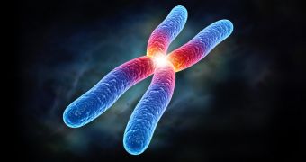
Scientists led by Dr. Peter Fraser of the Babraham Institute have managed to piece together the first ever 3D portrait of chromosomes.
The researchers say that, as this portrait shows, the X-shaped blob of DNA that most people are all too familiar with is by no means an accurate image of a chromosome.
In a paper recently published in the journal Nature, the scientists argue that, according to their investigations, chromosomes are only shaped like an X when the cell they sit in is about to divide.
Once the division process comes to an end, their shape is a much more complex one.
“The image of a chromosome, an X-shaped blob of DNA, is familiar to many but this microscopic portrait of a chromosome actually shows a structure that occurs only transiently in cells – at a point when they are just about to divide,” Dr. Peter Fraser explains, as cited by EurekAlert.
“The vast majority of cells in an organism have finished dividing and their chromosomes don't look anything like the X-shape. Chromosomes in these cells exist in a very different form,” the scientist goes on to say.
Apparently, the run-off-the-mill chromosome looks more like intricate folds of DNA. Its appearance depends on which genes are expressed.

 14 DAY TRIAL //