Doctors could soon have access to an improved understanding of biological process, thanks to technologies being created to measure the mechanical properties of living cells. This branch of mechanics has been studied only marginally thus far, but that will soon change.
Biologists have largely ignored the role that cellular mechanics play in the way cells react to, or influence, their environment and neighbors. Their interactions with the outside world, and even the process that go on inside, have until now only been analyzed from a chemical perspective.
However, it could be that some of their most important capabilities are made possible by some type of movement or another; and researchers have no way of knowing that at this point. But Purdue University experts are taking important strides in ensuring this lack of knowledge is addressed.
Purdue professor of mechanical engineering and team leader Arvind Raman explains that his group used an atomic force microscope (AFM) to study three different types of cells. Through this approach, the experts were able to demonstrate the versatility of using AFM for such investigations.
“There's been a growing realization of the role of mechanics in cell biology and indeed a lot of effort in building models to explain how cells feel, respond and communicate mechanically both in health and disease,” researcher Sonia Contera explains.
“With this paper, we provide a tool to start addressing some of these questions quantitatively: This is a big step,” she adds. The expert is an academic fellow at the University of Oxford Department of Physics, the director of the Oxford Martin Program on Nanotechnology, and a coauthor of the paper.
The joint Purdue/Oxford group published all details of its research in the latest online issue of the top scientific journal Nature Nanotechnology. The findings will also appear in the December print issue of the same magazine.
What the AFM-based study method provides is the ability to monitor cells as they are under the influence of drugs, antibiotics, alcohol, nicotine and other dangerous or helpful chemicals. This will allow experts to track any deformations that may occur as a result of exposure to these substances.
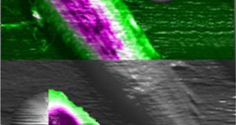
“The maps identify the mechanical properties of different parts of a cell, whether they are soft or rigid or squishy. The key point is that now we can do it at high resolution and higher speed than conventional techniques,” Raman explains.
He adds that funds for the new study were secured from the US National Science Foundation (NSF) and the UK Engineering and Physical Sciences Research Council (EPSRC).

 14 DAY TRIAL //