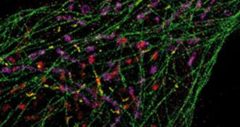
Investigators with the Wyss Institute of Biologically Inspired Engineering at the Harvard University announce the development of a new imaging method that enables the use of microscopy to identify individual biomolecules within living human cells.
The approach can create snapshots of multiple biomolecules at once within the same cell, without damaging or killing the latter. Details of how the technique works were published in the February 9 issue of the top scientific journal Nature Methods.
Researchers with the Harvard team say that having this capability could enable doctors to track the progress of diseases much more accurately. Furthermore, by analyzing the complex molecular pathways at work inside cells, doctors will be able to put relevant diagnostics quicker than ever.
“Peng's exciting new imaging work gives biologists an important new tool to understand how multiple cellular components work together in complex pathways. I expect insights from those experiments to lead to new ways to diagnose and monitor disease,” comments Don Ingber, the director of the Wyss Institute.

 14 DAY TRIAL //