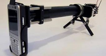
Most people use the cameras on their cell phones to capture instant moments while out with their families, or to photograph fun times with their pets. But experts from the University of California, in Berkeley, have other plans with their mobile phones. Their newly developed technology, CellScope, allows for average cell cameras to be retrofitted with powerful microscopes, able to detect malaria parasites, and even fluorescent marker-stained tuberculosis bacteria.
The new instrument could open the way for a new type of investigation medicine that takes place outside the classical frame of the science laboratory, the UCB team believes. Because of the high mobility of these devices, they can be transported virtually anywhere, and used on the spot wherever suspicions of contamination exist. The prototype CellScope device is described in detail in the latest issue of the scientific journal PLoS One, a publication of the Public Library of Science.
“The same regions of the world that lack access to adequate health facilities are, paradoxically, well-served by mobile phone networks. We can take advantage of these mobile networks to bring low-cost, easy-to-use lab equipment out to more remote settings,” Dan Fletcher, who is an associate professor of bioengineering at UCB, says. He has also been the leader of the science team that has developed the new CellScope prototype.
“The images can either be analyzed on site or wirelessly transmitted to clinical centers for remote diagnosis. The system could be used to help provide early warning of outbreaks by shortening the time needed to screen, diagnose and treat infectious diseases,” University of California in San Francisco (UCSF)/UCB Bioengineering Graduate Group graduate student David Breslauer adds. He has also been the co-lead author of the new paper.
The scientists also reveal the fact that the complex requirements of fluorescence microscopy, a highly complex procedure in the lab, have been met using nothing more than a cell phone camera and some inexpensive components that are readily available in stores. The new system employs several built-in filters, which ensure that background light from near the sample is blocked, as well as LEDs able to incite reactions in the markers placed in TB-infected blood samples.
“We had to disabuse ourselves of the notion that we needed to spend many thousands on a mercury arc lamp and high-sensitivity camera to get a meaningful image. We found that a high-powered LED – which retails for just a few dollars – coupled with a typical camera phone could produce a clinical quality image sufficient for our goal of detecting in a field setting some of the most common diseases in the developing world,” Fletcher concludes, quoted by ScienceDaily.

 14 DAY TRIAL //