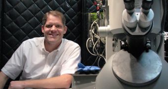
Cell division, the process through which a cell produces an exact copy of itself, can still baffle experts decades after the first studies began. In a recent investigation, a team of scientists discovered that one of the characteristic steps of mitosis is significantly different in some cells than in others.
California Institute of Technology (Caltech) researchers used a combination of powerful medical imaging technologies and cell sample slicing methods in order to observe the cellular nucleus while cell division was occurring.
The steps this process follows are very well understood, but there are some blind spots as well. For example, there are structures and molecules of certain dimensions in the cell that cannot be spotted by any microscope for a simple reason.
Modern equipment can focus on either the surface of the cell, or its most intimate innards, but not in between the two. A lot of effort is currently being expended to develop imaging technologies that would cover the gap, finally revealing the entire mechanism.
The Caltech team focused on mitosis because this stage of cell division leads to the pairing up of two sets of chromosomes at the center of the nucleus. The spindle apparatus in the cell then captures these structures, pulling them in opposite directions.
The apparatus is made up of microtubules, hollow-rod like protein structures without which mitosis would be impossible. These formations are in a way responsible for ensuring that each cell copy resulting from division carries a full, unadulterated copy of the original genetic material.
Before the split, each chromosome is grappled by one or more microtubules. At least, this is what experts thought after countless studies on plants, fungi and other species. However, the new observation sessions revealed an interesting difference.
“We've found the first clear example of a cell where there are fewer microtubules used than chromosomes,” explains Howard Hughes Medical Institute investigator Grant Jensen, who is also a professor of biology at Caltech.
Details of the new research effort appeared in the latest online issue of the esteemed scientific journal Current Biology, and will be published in the September 27 print issue of the magazine as well.
The US National Institutes of Health, the Gordon and Betty Moore Foundation, and a fellowship from the Damon Runyon Cancer Research Foundation provided the funds needed for this research.

 14 DAY TRIAL //