14 DAY TRIAL //
 It is said that our brain can organize itself only during our childhood, when it forms connections and pathways between different groups of neurons (brain cells that form the gray matter of the brain). The plasticity of the young brains enables them to change or adapt. This is how children struck by polio can still walk afterwards.
But a new case on a stroke patient points that adults' brains could be as plastic (capable of making new neural pathways) as those of children. The visual center of the stroke patient reorganized itself to overcome pathways destroyed by the stroke, improving changes in visual perception.
A team led by Dr. Daniel Dilks, while a graduate student at Johns Hopkins University, investigated the brain of a stroke patient, referred to as BL.
BL's stroke had harmed the fibers sending data from the eyes to the visual cortex, in the back of the brain. The cortex itself was untouched by the stroke. As a consequence, the connection between the upper left visual field and the area of the visual cortex where it projected its information was cut off, resulting a blind area in that upper left visual field. (A visual field means the area that can be seen when that eye is headed forward, including the peripheral vision.)
BL said things "looked distorted" in the area just below the impaired spot. The team presumed the distortions were caused by reorganization in the deprived cortex.
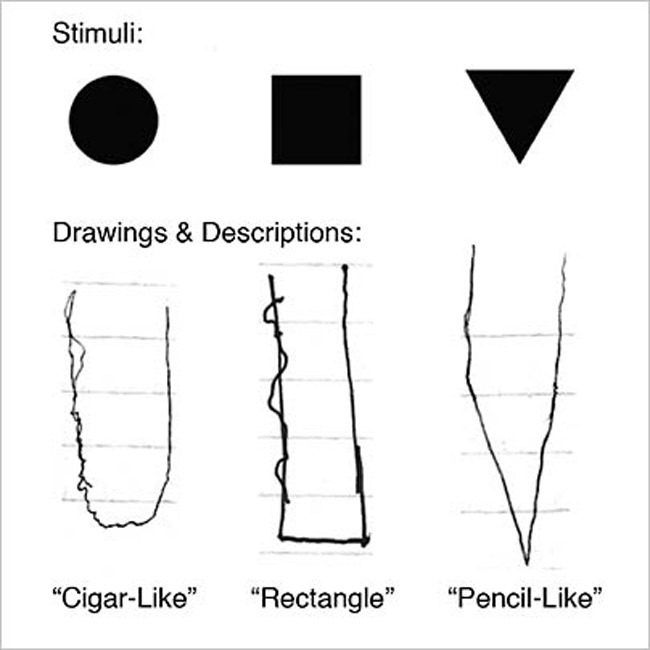
To check it, neuroscientists put BL to focus on a center dot while images of objects, like square shapes, appeared in different areas of the visual field. When the square was located in the blind area, BL saw nothing, but below the blind zone, BL saw the square as a rectangle stretching upward into the impaired area. BL detected triangles as "pencil-like," and circles as "cigar-like."
fMRI brain scans revealed that the visually impaired cortex (upper left visual field) was receiving optic information from the lower left visual field, a phenomenon not encountered in a healthy adult brain.
"That ability to "redirect" sight signals is a hallmark of plasticity and could explain the visual distortions." wrote the authors. "We discovered that it took on new functional properties, and BL sees differently as a consequence of that cortical reorganization," said Dilks.
Other researches, too, pointed out that there is a capacity of the adult brains to change. A 2007 research found adult mice could produce new neurons, with implications for the treatment of human neurodegenerative conditions like Parkinson's and Alzheimer's. A 2005 human adult brain-scan research on individuals with macular degeneration (which causes blindness in elders) found proofs of plasticity in the brain's visual areas.
Still, other brain scan researches failed to discover proof for brain plasticity in cases of macular degeneration.