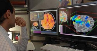
According to the conclusions of a new study conducted by researchers at the University of Michigan, it would appear that the very structure of the human brain is significantly affected by pain pill intake. The work suggests that this type of medication should be prescribed more carefully.
For this research, scientists used three different, high-resolution brain imaging techniques, in order to get the best possible data. This is the first study to look at the interactions that occur between compounds in pain medication and various types of cells in the human brain.
The investigation was focused on a drug usually prescribed to people suffering from fibromyalgia and neuropathic pain, known by the brand name Lyrica. The active ingredient in this pill is a compound called pregablin, PsychCentral reports.
U-M scientists used functional magnetic resonance imaging (fMRI), functional connectivity magnetic resonance (fCMR) and proton magnetic resonance spectroscopy to look at the brains of 17 patients diagnosed with fibromyalgia, a condition characterized by chronic pain and sensitivity to pressure.
In addition to bringing about pain from no particular source, this condition also sets the stage for the development of a number of mood disorders, including depression and anxiety. Statistics show that it affects between 3 and 6 percent of the world's population, and 10 million people in the US alone.
The study revealed that pregablin reduces the concentration of glutamate in the insula, a region of the brain the processes pain and emotions. However, the team also noticed that the compound reduced the neural connectivity between the insula and other parts of the brain.
“The significance of this study is that it demonstrates that pharmacologic therapies for chronic pain can be studied with brain imaging,” explains U-M assistant professor of anesthesiology, Richard Harris, PhD, who was also the lead author of the research.
“The results could point to a future in which more targeted brain imaging approaches can be used during pharmacological treatment of chronic widespread pain, rather than the current trial-and-error approach,” he goes on to say.

 14 DAY TRIAL //