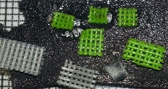
Vilnius University scientists Dr. Mangirdas Malinauskas (Laser Research Center) and Dr. Virginija Bukelskienė (Institute of Biochemistry) have introduced a scientific breakthrough that might pave the way towards a new age of (half-)synthetic implants.
Basically, they were able to combine three major techniques and eventually create hard tissue implants that can meld with and/or be absorbed by the body over time.
Implants, as they are now, are all well and good, but they don't usually restore a person's health completely, especially when the range of motion of a limb or other was affected.
3D printing technology allows for very precise reproduction of the natural bone however (or whatever else) and can lead to more highly biocompatible implants (when using bioplastics or stem cells).
As it happens, stem cells definitely played an important role in the latest scientific breakthrough.
Three techniques were combined to achieve a new implant production method
In essence, fused filament fabrication (FFF) 3D printing (otherwise known as fused deposition modeling, or FDM) was combined with direct laser writing (DLW) and 3D bioprinting.
Which is to say, while the scaffolds were made of PLA, they were seeded with stem cells, rabbit stem cells to be exact.
Tests later showed that the resulting technology combo can be used to produce highly effective and safe hard tissue implants, soft tissue treatments, and basically a whole slew of other biomedical surgery techniques.
It all sounds impressive, because it is, but what makes this breakthrough really astonishing is that it was achieved using a normal Ultimaker 3D printer, the sort even you should be able to buy for a few hundred bucks.
Then again, you do need an amplified femtosecond laser to create microstructures, and the acetone bath that follows is necessary to boost the hydrophilic properties of the whole thing (nanoscale roughness), otherwise cells won't adhere to it very well.
After an electron microscope scans the scaffolds, stem cells are seeded and tested for viability via a fluorescence microscope.
The end result is an implant (the scaffolds, basically) that can be implanted and left in the body, where it will decompose in 2 years or so. More than enough time for cells to grow through the constructs and eventually replace the hard tissue altogether.
Other applications of the technique
Components in microfluidics and micromechanics should be easy enough to make this way, since the filling ratio (porosity) of a surface area can be very well controlled by the scientists / mechanics / engineers / scientists.

 14 DAY TRIAL //