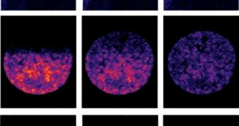
Scientists in Europe recently managed to perfect a new achievement in the field of microscopy, when they improved the functionality of a software program that enables microscopes to “learn” what their users are searching for in images.
This allows for the machines to conduct studies autonomously, and pinpoint the location of specific structures inside cells, that are similar to structures researchers gave as a reference point.
The German research team that developed the software was capable of using it to perform a series of complex microscopy experiments, and then published those results in the latest issue of the esteemed scientific journal Nature Methods.
At this point, when researchers want to find something in a sample, such as for example a specific type of cell, they need to sit over the microscope for hours, manually searching for what they need to see.
But this software, developed by experts at the European Molecular Biology Laboratory (EMBL), in Heidelberg, could make that a thing of the past. The program will learn what each researcher is trying to find, and will conduct the search on its own.
This will free up investigators to focus their attention on something else rather than the tedious microscope search. In addition, the software can also tell the microscope to conduct advanced studies of samples, if they contain cells that are deemed of interest.
The basis of the Micropilot (microscope pilot) software is combining the field of microscopy research with machine learning and advanced artificial intelligence (AI). The program controlling the scientific tool only tells the microscope to start studies once interesting features are identified.
This is done after Micropilot analyzes low-resolution images of the samples. Its algorithms can easily detect the existence of interesting structures, or components of the type that have been observed by researchers before, AlphaGalileo reports.
The experiments the software can conduct range from recording high-resolution, time-lapse videos of various cellular components in action to using lasers to disrupt the behavior of fluorescent proteins and analyzing what happens.
In experiments meant to assess the new technology, the EMBL team left Micropilot unattended for four nights. During this time, the program identified 232 cells in two particular stages of cell division, that the team was interested in.
In addition, the program instructed the microscope to perform a complex imaging experiment on these cells that generated a wealth of data. The real kicker is that a human operator would have needed at least a month to find the 232 cells, the EMBL team says.

 14 DAY TRIAL //