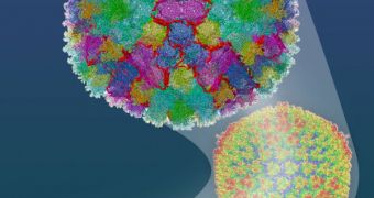
For years, researchers have been unable to image the viruses they were trying so hard to destroy, but now not only has that become possible, but they can also use the microorganisms to deliver drugs.
This is possible thanks to efforts by a team of investigators based at the University of California in los Angeles (UCLA).
The group was led by expert Hongrong Liu, who is a postdoctoral researcher in microbiology, immunology and molecular genetics at the university.
The scientist and his group published the full details of their accomplishment in last week's issue of the esteemed journal Science.
What the UCLA multidisciplinary research group did was basically develop a method of visualizing viruses, while at the same time allowing investigators to manipulate them so that they can carry drugs.
As such, rather than spreading disease, the microorganisms begin spreading the cure for whatever disease is needed. Or, at least, this is the hope the UCLA team has in store for the adenoviruses.
“We are engineering viruses to deliver gene therapy for prostate and breast cancers, but previous microscopy techniques were unable to visualize the adapted viruses,” explains Lily Wu.
“This was like trying to a piece together the components of a car in the dark, where the only way to see if you did it correctly was to try and turn the car on,” adds the expert, who is a UCLA professor of molecular and medical pharmacology.
She was also the co-lead author of the new journal entry. Wu explains that, until now, a crippling lack of knowledge about receptors on the virus's surface had hampered research into this issue.
Gaining new insights into how the viruses looked like meant creating atomically-accurate, three-dimensional models of biological samples.
This why the research team enlisted the help of UCLA professor of microbiology, immunology and molecular genetics Hong Zhou, the other co-lead author of the Science paper.
The expert was able to produce the required models using an observations technique called electron microscopy (cryoEM)
“Because the reconstruction reveals details up to a resolution of 3.6 angstroms, we are able to build an atomic model of the entire virus, showing precisely how the viral proteins all fit together and interact,” Zhou reveals.
Funding for the new investigation came from the US National Cancer Institute (NCI) and the Department of Defense (DOD).

 14 DAY TRIAL //