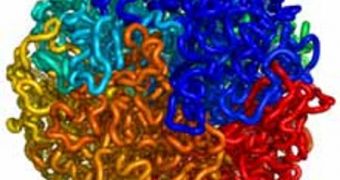
As most of you know, unfurling the genetic material enclosed in each of our cells would result in a six-foot-long strand of DNA. However, inside each cell, all this information remains stored within nuclei that are less than three micrometers in diameter, less than the width of a human hair. Finding out precisely how these molecules are crammed into such a tiny space has been something experts have been working on for a long time, and it would appear that the new investigation method has now finally discovered the answer, Technology Review reports.
The method can determine the three-dimensional interactions that take place between the various parts of the genome, essentially hinting at how the genetic material and DNA molecules pack themselves inside the nuclei. Previous studies have already determined the 3D structure of several parts of the genome, but the newly developed instrument is the first to do so on a genome-wide scale, its creators say. “Our technology is kind of like MRI for genomes,” Harvard-MIT Division of Health Sciences and Technology researcher Erez Lieberman-Aiden explains. The scientist is also one of the authors of a new paper detailing the find.
The thing about DNA is that it not only organizes itself in linear strands and the basic double-helix structures, but also in higher-order structures, when it wraps around the proteins and triggers the formation of chromosomes. It's precisely this higher-order levels that have largely remained a mystery, and that are now the target of this new method. “We have the entire linear sequence of the genome, but no one knows even the principles of how DNA is organized in higher-order space,” National Cancer Institute (NCI) expert Tom Misteli, who has not been part of the new study, explains.
The new method, dubbed Hi-C, is fairly complex. First, formaldehyde or some other preservative is used to fix a complex DNA structure in place. Enzymes are applied to the mix, which break the formation into thousands of smaller pieces, save for the bits of DNA that have been treated with the preservatives. At the sites where the bonded fragments end, experts then place a substance called a biotin, and use another type of enzyme to bring two or more of the fragments together. This results in a circle of DNA, the team explains. All the pieces marked with biotin are then sequenced, and they reveal the original, 3D structure from which they came.

 14 DAY TRIAL //